Wednesday, May 21, 2014

Lab Day 5

Upon entering lab, we checked yesterday’s experiments. In our thioglycolate slants and we found growth on the top and the bottom, therefore signifying that our bacteria is a facilitative aerobe. Our stab was not motile because it remained in the line of inoculation and was not hazy throughout the tube. Our plate had growth and this further confirmed that it was a facilitative aerobe.

The second part of our lab consisted of seeing if our unknown bacteria had catalase. Any oxygen breathing organism can produce H2O2 because they contain the catalyse enzyme which breaks down H2O2 into Hydrogen and Oxygen. By pouring Hydrogen Peroxide onto the plate with our unknown bacteria, we could determine if our bacteria did in fact have cataylse. There were bubbles around the reaction which means our bacteria tested positive for cataylse. We are O2 breathing beings, therefore, also have catalyse and can produce H2O2.
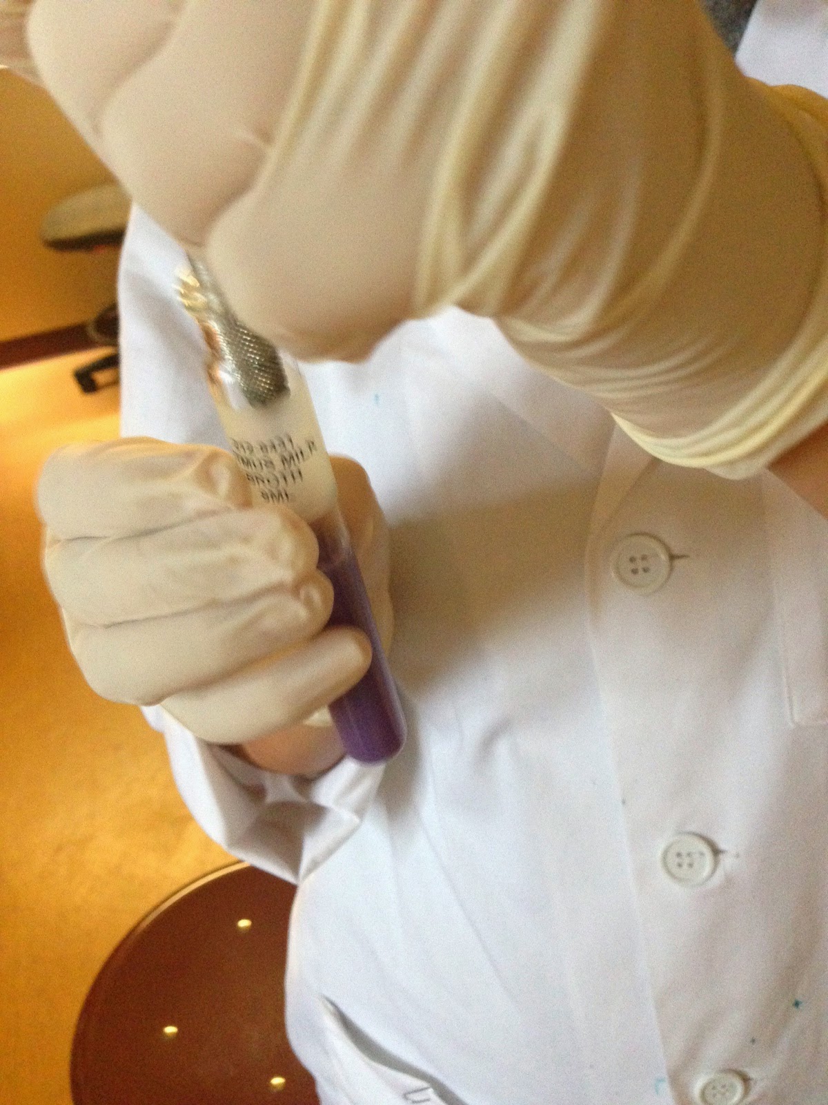

 During the last part of our lab, we prepared tests that would help us discover what foods our bacteria prefer to eat such as lipids, proteins, carbohydrates, or nucleic acids. To do this, we used the aseptic technique to inoculate many test tubes with different foods such as glucose, sucrose, maltose, lactose, and litmus milk, with our unknown bacteria. We took a test tube containing TSIA and stabbed our bacteria into the slant and then used the snaking technique over the surface of the slant. We also inoculated a starch agar with our bacteria. After 24 hours you stain the plate with Gram's Iodine. We inoculated a skim milk agar using the snaking method. The last two agar plates we inoculated were the DNA and Lipid plates.


Joanna Dawyot, Cassie Livingston, Mary Rose Capara 

DisclaimerAll content provided on this blog is representation of the blog owner and not FranciscanUniversity of Steubenville. The information on this site is purely used for education purpose. The owner of this blog makes no representations as to the accuracy or completeness of any information on this site or found by following any link on this site. The owner will not be liable for any errors or omissions in this information nor for the availability of this information. The owner will not be liable for any losses, injuries, or damages from the display or use of this information. Privacy The owner of this blog does not share personal information with third-parties nor does the owner store information is collected about your visit for use other than to analyze content performance through the use of cookies, which you can turn off at anytime by modifying your Internet
 browser’s settings. The owner is not responsible for the republishing of the content found on this
 blog on other Web sites or media without permission.Blog CommentsThe owner of this blog reserves the right to edit or delete any comments submitted to this blog without notice due to;1. Comments deemed to be spam or questionable spam2. Comments including profanity3. Comments containing language or concepts that could be deemed offensive4. Comments that attack a person individuallyThis policy is subject to change at anytime.

Tuesday, May 20, 2014

Sick Days and Bacteria Tests

Day 4
May 19, 2014

Today was the day we all found out about our acceptance into the nursing program!! Lab began with Dr. P teasing Elizabeth about her acceptance. She was afraid she didn't get in and didn't want to get her letter from Dr. Miller but in the end, we got her to smile with the good news.
Saturday night, Pete, Dr. P, Mary Rose, Ben, and Adrianna went to the lab to take a culture of Mary Rose's throat, thinking that she had strep. The plate with the swab had incubated over two nights and we were ready to see the results.
The plate had obvious bacterial growth and as it turns out, Mary Rose tested positive for strep. Pete had experimented with the strep by adding Penicillin, cinnamon oil, and clove oil to the plate. We were able to link lecture to lab by observing the changes in the bacteria. The cinnamon oil killed RBC but did the best job of killing the strep. It did a better job at this than Penicillin! Clove oil also did an incredible job at killing the bacteria as did Penicillin. We were able to deduce the bacteria's death due to clear rings around each of the three substances on the plate.
Now we know! If you have strep or a throat infection, take cinnamon and clove!!

Part 1: Thioglycolate

Practicing Ascetic technique throughout the experiment, we took a loop and, taking a culture of our unknown bacteria, we dipped it into a test tube filled with thioglycolate. This substance was pink towards the top of the tube and a yellowish color on the bottom. This was due to the thioglycolate being oxygenated on the top and not the bottom. By stabbing our bacteria into this tube, we will be able to see if our bacteria is aerobic or anaerobic due to how it will grow in the incubator overnight.
Upon receiving Dr. P's permission, we repeated this procedure with Mary Rose's strep culture. This too we put into the incubator.

Part 2: Stab to test Motility

This we did by taking our loop with our unknown bacteria and stabbing it into an agar deep provided by Dr. Pathakamuri. We put it into the incubator and we'll see what happens tomorrow!

Part 3: Inoculation of a Slant Agar

Because our first slant samples melted we had to redo the two agar slant samples we did earlier last week. Taking our bacteria with a loop, and practicing ascetic technique throughout, we used the snake method to spread the bacteria on the surface of an agar slant. We'll see what happens to the bacteria tomorrow!

Part 4: Testing the effects of a virus on bacteria

We began this procedure by spreading 50ml of group 1's, our group, broth onto a plate. This we did by using a dispenser set to gather 50ml of the broth. Dr. Pathakamuri gave each of the students a small amount of an unknown virus and, taking a big tip, we each wrote something on the broth with it. Our group chose to write Europe! We're excited to be going officially now that we're                      in the program!
After that, we put the plates the incubator for viral growth.

Part 5: Culturing Anaerobic Bacteria

Taking a small sample of our bacteria, we spread this on a plate. We then put the plate into an anaerobic jar. This jar is used to test whether or not our bacteria is anaerobic. This jar expels all traces of oxygen by the use of hot gas packs in the air tight container. We also put an indicator strip into the box. This will indicate the presence or lack of oxygen. Pale white = no oxygen and blue = presence of oxygen.
We'll get all of our results tomorrow!!


Joanna Dawyot, Cassie Livingston, Mary Rose Capara 

DisclaimerAll content provided on this blog is representation of the blog owner and not FranciscanUniversity of Steubenville. The information on this site is purely used for education purpose. The owner of this blog makes no representations as to the accuracy or completeness of any information on this site or found by following any link on this site. The owner will not be liable for any errors or omissions in this information nor for the availability of this information. The owner will not be liable for any losses, injuries, or damages from the display or use of this information. Privacy The owner of this blog does not share personal information with third-parties nor does the owner store information is collected about your visit for use other than to analyze content performance through the use of cookies, which you can turn off at anytime by modifying your Internet
 browser’s settings. The owner is not responsible for the republishing of the content found on this
 blog on other Web sites or media without permission.Blog CommentsThe owner of this blog reserves the right to edit or delete any comments submitted to this blog without notice due to;1. Comments deemed to be spam or questionable spam2. Comments including profanity3. Comments containing language or concepts that could be deemed offensive4. Comments that attack a person individuallyThis policy is subject to change at anytime.


Friday, May 16, 2014

Staining Day!!

Lab Day 3
“I was Born Ready” -PiCassie

Today's lab began by Mary Rose spraining her ankle playing ultimate frisbee. Woohoo. After being brought into the lab in a wheelchair, Joanna and Cassie joined to begin today's procedures.

Part 1 of today's lab: Gram Stain

When we started preparing to do the gram stain, everyone realized that we, the class, had left our samples in the incubator instead of the refrigerator. According to Dr. P, “One night in there is more than enough.” Oops! To our relief however, no harm had been done to our samples.
Using ascetic technique, we made a fixed smear of our bacteria. We then applied Crystal Violet dye on the air-dried sample for 20 seconds. Using bibulous paper, we pat dry the slide so no excess water would interfere with our observations under the microscope. After this, we applied the Gram Stain and let is sit for 60 seconds. Whoever had the idea to smell this (Mary Rose) made a mistake. It smelled like nasty latex. 

Lab was interrupted by a surprise visit from Dr. Pathakamuri's family which brightened everyone's mood. Dr. P jokingly asked his youngest son to help us look into the microscope but he quickly refused. To remove the Gram Stain, we added 95% Ethanol until the color on the slide stopped running. After this, we applied Gram Safranin, which is a reddish color, also for 60 seconds. We then observed the slide under the microscope and, with the help of Dr. Pathakamuri, we concluded that our unknown bacteria was gram negative and rod shaped. With that knowledge, we knew that our bacteria had a thinner cell wall and thinner peptioglycan layer and that of a gram negative cell wall.

Part 2 of today's lab: Capsule stain

The next experiment we preformed on our unknown bacteria was a Capsule Stain. First, we placed a small amount of Nigrosin dye on a slide (slide #1) with a sample of our unknown bacteria. We then mixed the two together and, using the edge of another slide (slide #2), smeared the dyed bacteria mixture across the surface of slide #1. After the smear dried, we covered it with the crystal-violet dye and let it sit for a minute over the sink. That's when Pathakamuri accidentally knocked a slide over that proceeded to shatter on the ground. Everyone laughed when he turned to Professor Lumbar and said, “Professor Lumbar, someone left a depression slide out and it wasn't one of my students. I don't know whose fault that is!”
Now we know how to handle the situation if we ever break a slide!
We washed off the excess dye with DI H2O but we did not do this for a long period of time due to the possibility of disrupting the placement of the original smear. We then blotted the slide with the bibulous paper and headed over to the microscope. There, we were able to see that our unknown bacteria has no capsule.


Part 3 of today's lab: Acid-fast Stain
Before beginning our final experiment of the day, the acid-fast stain, Dr. Pathakamuri put music on the surround speakers in the lab. This turned out to be one of our favorite memories from lab thus far. We listened to Enya, the Lion King, Dr. Pathakamuri's choir Cd, and various other types of music. That's when we realized that Pete and Cassie have incredible opera voices! (insert video)
We prepared another fixed smear and placed it over a beaker of boiling water. We applied Carbolfuchsin, a green dye, onto a piece of bibulous covering the slide and let it sit for 5 minutes. We were careful to not let the dye dry out by continually saturating the paper with excess Carbolfuchsin.
When 5 minutes was up, we removed the slide from the beaker with thongs and discarded the piece of paper in hazardous waste bin. We let the slide cool and then rinsed it with water to remove excess stain. We decolorized the slide with acid-alcohol while holding the slide at a 45 degree angle. We did this until the color stopped running then immediately rinsed the slide to remove the decolorizing agent.
We covered the smear with Methylene blue and let that dye sit for 2 minutes. We rinsed the stain off with water to remove excess dye and observed it under the microscope. We repeated this same procedure for an unknown bacteria given to us by Dr. Pathakamuri.

The results:

  Our unknown bacteria was negative for being an acid-fast bacteria and Dr. Pathakamuri's unknown was positive for being an acid-fast bacteria.


Part 4 of today's lab: Endospore Staining

This staining for our unknown sample showed that our bacteria had no endospores present. 


End of the Lab Day Results!!!


Joanna Dawyot, Cassie Livingston, Mary Rose Capara 

DisclaimerAll content provided on this blog is representation of the blog owner and not FranciscanUniversity of Steubenville. The information on this site is purely used for education purpose. The owner of this blog makes no representations as to the accuracy or completeness of any information on this site or found by following any link on this site. The owner will not be liable for any errors or omissions in this information nor for the availability of this information. The owner will not be liable for any losses, injuries, or damages from the display or use of this information. Privacy The owner of this blog does not share personal information with third-parties nor does the owner store information is collected about your visit for use other than to analyze content performance through the use of cookies, which you can turn off at anytime by modifying your Internet
 browser’s settings. The owner is not responsible for the republishing of the content found on this
 blog on other Web sites or media without permission.Blog CommentsThe owner of this blog reserves the right to edit or delete any comments submitted to this blog without notice due to;1. Comments deemed to be spam or questionable spam2. Comments including profanity3. Comments containing language or concepts that could be deemed offensive4. Comments that attack a person individuallyThis policy is subject to change at anytime.


Thursday, May 15, 2014

Day 2 In the Journey to New Discoveries
May 15, 2014

Being that we had a quiz in lecture, all of us were feeling a bit tired and overwhelmed from learning, what we felt was, everything a person could possibly know about viruses. Regardless, we were excited and curious to see what growth our unknown bacteria underwent.

Part 1 of Today's Lab: Observing Growth
After being instructed to remove our sample bacteria slants, plate, and broth from the 37 degree Celsius incubator, we sat down to examine the changes they had undergone. With simply a naked eye, it was obvious the bacteria had grown.
    1. The Plate
      The plate had the most obvious growth of bacteria out of our three different bacteria samples. Under microscopic view, we were able to see that multiple single colonies had emerged along the boarder. According to Dr. P, our bacteria was “a fast growing one.” The visible characteristics of the bacteria are as follows:
               Colony Pigmentation: cream/tan

      Whole Colony (form): circular

      Colony Margin (edge): undulate

      Elevation of Colony: hilly


                 II.The Broth
While it wasn't obvious at first glance that the bacteria had grown overnight, we performed a flicking motion against the bottom of the test tube to stir up the bacteria sediment. The broth then became more cloudy/hazy.


                 III.The Agar Slant
 Observing the bacteria growth both with the naked eye and under the microscope, we were able to deduce that the bacteria growth on the agar slant was beaded.


Part 2 of today's lab: Observing the Motility of Our Unknown Bacteria
Being that our bacteria had growth, we then prepared, still tired but certainly interested, to see if our bacteria was alive and moving.
Materials used:

Cover Piece
Bunsen Burner
Loop Tool
Depression Slide
Microscope 
Unknown Bacteria
Vaseline 


Procedure:
   Using a loop and of course, aseptic technique, we took a sample of bacteria from our broth and after flicking the bottom to ensure a well mixed sample, we placed a drop on a cover piece provided. After adding vaseline to the corners, we attached it to the depression slide and flipped it over. This type of experiment is known as the Hanging Drop Experiment.


With a compound microscope, we decreased the light completely to ensure visibility of the bacteria. Between our compound microscope and Dr. Pathakamuri's microscope, we were able to see that our bacteria was swimming around.


Dr. P suggested that we add broth to the cover slide to further observe the bacteria's motility. The projected image of the microscope's view revealed that the bacteria seemed to “stand up” in attempts of broth consumption.


Part 3 (our favorite part) of today's lab: Simple Staining
Following our observation of the bacteria's motility, we were instructed by Dr. P to begin a simple stain experiment on the bacteria.

Materials Used:

Slide
Loop
Bunsen Burner
Crystal Violet dye
DI H2O
Tongs
Microscope
Bibulous Paper

Procedure:
We began by cleaning the slide with a task wipe and, practicing aseptic technique throughout, we obtained a sample of our bacteria from our plate and spread it into a thin film on the slide. We allowed it to air dry in order that the moisture in the sample evaporate. Then, using tongs, we swiftly passed in through a flame, bacteria side facing up, three times. The bacteria was then considered “heat-fixed.”


 We covered the fixed smear with several drops of crystal violet stain and let is sit for 25 seconds, as directed by the lab manual. Then, we rinsed the slide with DI H2O to remove excess stain and blotted the water with pieces of bibulous paper.


After that, we examined the newly stained bacteria under a blue light to discover the shape of the bacteria.
Under the lowest magnification, it appeared that our bacteria was round. However, under the highest magnification, we discovered that our group's bacteria had groups of small rods.


This concluded day 2. We look forward to seeing new growth tomorrow and experimenting to discover our bacteria's identity .

Joanna Dawyot, Cassie Livingston, Mary Rose Capara 

DisclaimerAll content provided on this blog is representation of the blog owner and not FranciscanUniversity of Steubenville. The information on this site is purely used for education purpose. The owner of this blog makes no representations as to the accuracy or completeness of any information on this site or found by following any link on this site. The owner will not be liable for any errors or omissions in this information nor for the availability of this information. The owner will not be liable for any losses, injuries, or damages from the display or use of this information. Privacy The owner of this blog does not share personal information with third-parties nor does the owner store information is collected about your visit for use other than to analyze content performance through the use of cookies, which you can turn off at anytime by modifying your Internet
 browser’s settings. The owner is not responsible for the republishing of the content found on this
 blog on other Web sites or media without permission.Blog CommentsThe owner of this blog reserves the right to edit or delete any comments submitted to this blog without notice due to;1. Comments deemed to be spam or questionable spam2. Comments including profanity3. Comments containing language or concepts that could be deemed offensive4. Comments that attack a person individuallyThis policy is subject to change at anytime.

Wednesday, May 14, 2014

Our Trip to the Gym Turns Scientific and Inoculation of Bacteria

Our Trip to the Gym Turns Scientific and Inoculation of Bacteria
May 14, 2014

Have you ever noticed the signs at the gym saying “Wipe down your machines once you are done with them”? When studying Medical Microbiology, Dr. Pathakumuri asked us to find a sample of bacteria on the campus. We took this opportunity to help people understand why wiping down machinery is so important.
We left the lab in our white lab coats ready to obtain our specimen. Upon entering the gym, we went to the most commonly used piece of equipment, the squat bar. To collect our sample, we practiced aseptic technique, using an agar broth and a cotton swab. Then we transferred the bacteria to an agar plate using the streak method, rotating the plate a quarter around every time we streaked the bacteria. Once we arrived back to the lab we placed the plate in an incubator that was set at 24 degrees C.


Inoculation of Bacteria.
Hypothesis: We believe that our unknown sample of bacteria that Dr. P gave to us affects the body in some way, being that the human body temperature is 37 degrees C as is the sample bacteria that we were given. Whether it has a beneficial purpose or detrimental affect, we are unsure of at the time being.
Our Goal: To make a pure stock.

Materials Used:
Loop
2 Agar Slant Tubes
Agar Plate
Bunsen Burner
Agar Broth
Gloves
Documentation Device

A. Transferring bacteria from a slant agar to a plate
Taking the loop we passed it through the tip of the inner cone of the flame to ensure sterilization. After we passed the mouth of the tube through the flame we dipped the loop into the bacteria sample. Then we used quadrant technique and the streak method, we spread the bacteria on the plate. After we finished each quadrant we sterilized the loop and then streaked the bacteria from the previous quadrant into the next one. This method ensures that we will grow a pure culture.


B. Transferring bacteria from a slant agar to a slant agar
Practicing aseptic technique throughout, we used the loop to transfer bacteria from one slant to the other. We did a shallow stab to collect the bacteria from the contaminated sample. Using a snake-like motion, we spread the bacteria onto the pure slant. We repeated this technique twice.


  1. Transferring bacteria from a slant agar to agar broth Practicing aseptic technique, we used the loop to transfer bacteria from the slant to the broth. We did another shallow stab to collect a small sample colony from the contaminated sample. We dipped the loop with the bacteria in to the broth.


                                          We then placed all 4 of the samples into an incubator at 37 degrees C.


Joanna Dawyot, Cassie Livingston, Mary Rose Capara 

DisclaimerAll content provided on this blog is representation of the blog owner and not FranciscanUniversity of Steubenville. The information on this site is purely used for education purpose. The owner of this blog makes no representations as to the accuracy or completeness of any information on this site or found by following any link on this site. The owner will not be liable for any errors or omissions in this information nor for the availability of this information. The owner will not be liable for any losses, injuries, or damages from the display or use of this information. Privacy The owner of this blog does not share personal information with third-parties nor does the owner store information is collected about your visit for use other than to analyze content performance through the use of cookies, which you can turn off at anytime by modifying your Internet
 browser’s settings. The owner is not responsible for the republishing of the content found on this
 blog on other Web sites or media without permission.Blog CommentsThe owner of this blog reserves the right to edit or delete any comments submitted to this blog without notice due to;1. Comments deemed to be spam or questionable spam2. Comments including profanity3. Comments containing language or concepts that could be deemed offensive4. Comments that attack a person individuallyThis policy is subject to change at anytime.